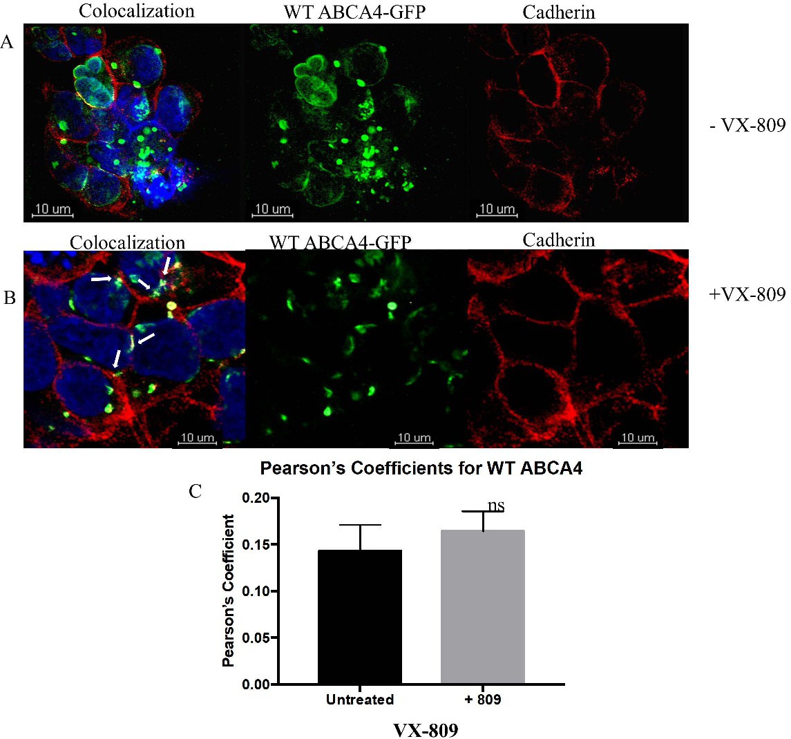
Fig. 7. Plasma membrane localization of WT-ABCA4 protein. (A) WT ABCA4-GFP (green) stably transfected cells were plated on coverslips. Cells were subsequently fixed and stained with DAPI (blue, nucleus) and antibodies against cadherin (red, plasma membrane). Areas of colocalization give off a yellow signal (white arrows). Representative extended focus images are shown. (B): Cell surface expression of ABCA4 protein is unaffected by VX-809. WT ABCA4-GFP (green) stably transfected cells were plated on coverslips. Cells were subsequently fixed and stained with DAPI (blue, nucleus) and antibodies against cadherin (purple, plasma membrane). Areas of colocalization give off a white signal (white arrows). 3 separate experiments were performed. Representative extended focus images are shown. (C): Pearson's coefficients were obtained for VX-809 treated and untreated cells. Coefficients were compared using the student's t-test and found to be non-significant (n=3).